")
")
")
Shoulder Center Saar
Needle Lavage: A New Therapy for Calcific Shoulder
While still largely unknown in Germany, ultrasound-assisted microinvasive lavage of calcified lesions in calcified tendinosis (calcified shoulder) has become established internationally. This procedure, which has been known for more than 10 years, has demonstrated its high success rate in several scientific studies. New, recently published studies even demonstrate success rates comparable to arthroscopic procedures for calcification removal.
If you are interested in this therapy, you can find further information here >>>The Development of the Procedure
The procedure of microinvasive ultrasound-assisted calcification removal is a further development of the needling of calcified shoulder lesions, which has been known for many years. Traditionally, an X-ray image intensifier is used for localization during needling. This device is a mobile X-ray machine that can not only take images but also film recordings in real time. After the patient is correctly positioned and the device is moved into position, a needle is inserted to target the calcification in the live X-ray image. The goal is to open the calcification and create a wound that will trigger the calcification to dissolve spontaneously.
Due to the two-dimensional imaging of the X-ray machine, the precise localization of the calcification is very difficult and can result in high radiation exposure for the patient.
Development boost through the use of modern ultrasound devices with 3D imaging
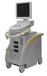
Through the use of new, high-resolution, modern "3D ultrasound technology," a calcification in a shoulder tendon can now be precisely located. The new ultrasound devices, with a resolution of 14 MHz, allow for unprecedented detail in the shoulder joint. After local anesthesia, a special needle is inserted through the skin. Continuous monitoring via live ultrasound allows the needle to be optimally placed in the calcification.
This technique for positioning needles or other instruments is not new, but is rarely used in orthopedics and surgery. In internal medicine, ultrasound-guided puncture is regularly used with great success for a wide variety of clinical pictures and for tumor diagnostics.
Once the irrigation needle is in the optimal position, the calcification is mechanically loosened by injecting a local anesthetic solution and then suctioned out. To remove as much calcium as possible from larger calcifications, the needle must be repositioned several times. Ideally, the calcium from the tendon is in the irrigation and suction syringe at the end of the therapy.
The major difference between microinvasive therapy and other therapies
In contrast to traditional needling, the calcification is flushed out using a cannula. This means that after the procedure, the suctioned calcification is placed in a syringe. With needling, the calcification remains in the tendon, and only a few holes are punctured in the calcification to encourage the calcification to dissolve spontaneously. Compared to shock wave treatment, there is a slightly increased risk, as this is a surgical procedure, albeit a very minor one. However, at the end of the treatment, the calcification is actually removed, rather than the hope of spontaneous dissolution of the calcification, as with shock wave therapy. Compared to arthroscopy, there is a significantly lower surgical and anesthetic risk, as the procedure is performed on an outpatient basis and under local anesthesia. According to current studies, the results are comparable in terms of success.
How is the treatment performed?
After an initial examination, in which the shoulder is again closely inspected using ultrasound, the arm is positioned to ensure optimal access to the calcification during the next steps. The surgical area is then carefully disinfected several times. Local anesthesia of the skin and the area surrounding the calcification is then administered under ultrasound guidance.
Next, as described above, a special irrigation cannula is advanced through the skin and placed into the calcification under careful ultrasound guidance. Once the position is optimal, the calcification is rinsed out. The following video explains the therapy again and demonstrates a treatment.
You must have the Adobe Flash Player installed to view this player.
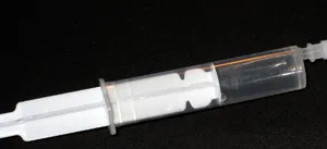
A local anesthetic is used to flush the calcification. Using this solution significantly reduces the pain experienced during treatment. The local anesthetic is clear as water. You can see a prepared syringe in the bottom left of the image. After the treatment, the calcium from the calcification is in the "flushing syringe" and settles at the bottom (bottom right of the image). In contrast to simple needling, this procedure involves true calcium removal. This is also the key difference from the long-established needling procedure.
Since questions about this therapy and its differences from needling have been increasing recently, you can find a detailed explanation under the "News" section of our website.
-
-
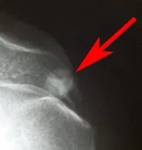
X-ray of a calcified shoulder at the start of treatment. The calcification between the acromion and the humeral head is clearly visible. The red arrow points to the calcification.
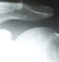
- On the right is the X-ray image 2 weeks after treatment. The calcification has almost completely disappeared.
You must have the Adobe Flash Player installed to view this player.
Since the rinsing process is also performed with a local anesthetic, the entire procedure is relatively painless. The procedure is performed on an outpatient basis, so the patient can leave our practice immediately afterward. Immediately after the calcium removal, the arm can be used again. Wearing the arm in a sling or immobilizing it is not necessary. After the procedure, symptoms usually improve significantly. In rare cases, increased discomfort occurs for a few days. This is caused by residual calcium, which dissolves further as part of a mild inflammatory reaction. However, this situation can also be easily managed with painkillers.
Overall, it often takes 6–8 weeks for the symptoms to completely subside. This is due to the hole in the tendon that remains after the calcification removal and must heal. To date, this procedure has only been performed in a few centers. The reason for this is the need to use a very high-resolution ultrasound device to reliably locate the needle position. Since these devices are extremely expensive, costing approximately 10 times as much as those commonly used in orthopedics, their availability is very limited. We are fortunate to have such an innovative device and to be one of the first practices in Germany to offer this new procedure.
We have been performing the procedure regularly in our practice since September 2007. The good prospects of success with minimal risk have led patients from far beyond the borders of Saarland to seek treatment with this modern procedure. Inquiries from neighboring European countries are also increasing. Several scientific studies demonstrate the excellent results of this procedure. Just a few days after the procedure, more than 50% of patients are virtually pain-free. Overall, the studies reported good or very good results in over 75% of cases. These results are consistent with our experience with this treatment procedure.
Comparing the current studies on shock wave therapy and microinvasive surgical therapy, microinvasive surgical therapy produces significantly better results in terms of calcification removal. For further information, we have provided several studies available for you to read in our download area.
Due to the positive results this procedure promises, the online magazine Focus Gesundheit also reported on the therapy we use. The study cites an Italian study published in the renowned scientific journal "Radiology" in the USA, which reported positive results in more than 200 patients. For more information, follow this link: Quick Help for Calcified Shoulder - Focus Gesundheit