")
")
")
Shoulder Center Saar
Shoulder Arthroscopy
Gentle Shoulder Therapy Thanks to Keyhole Surgery
Until recently, shoulder surgeries were complex and sometimes risky procedures. The results achieved were often only moderately satisfactory. In recent years, new minimally invasive surgical techniques have made it possible to treat many shoulder joint disorders that were previously impossible to treat or required extensive surgical intervention. Thanks to these new, joint- and tissue-sparing techniques, surgical successes are now possible that were unthinkable just a few years ago. The technique of arthroscopy (joint endoscopy) can now effectively help many patients.
How does a shoulder arthroscopy work?
Shoulder arthroscopy is a particularly joint-sparing surgical method. In contrast to open surgery with a single large incision, only two or three small incisions, each less than half a centimeter long, are required. A small rod camera with a connected light is inserted into the shoulder joint through one of these incisions. The camera transmits the image from the joint to a monitor, allowing the surgeon to see what the joint looks like from the inside. Instruments can be inserted into the joint through the other incisions to repair injured or diseased structures. For better visibility and to flush the joint, sterile fluid is also pumped into the joint. Inflammatory substances or bacteria that have entered the joint are immediately flushed out.
The advantages of shoulder arthroscopy
By using a magnifying lens, the experienced surgeon can precisely assess any damage to the shoulder joint and provide the optimal treatment. Unlike open surgery with large incisions, in which the shoulder joint is usually completely opened, arthroscopy requires incisions of only a few millimeters to access the damaged joint. This protects healthy structures that could potentially be damaged by a longer incision. Shoulder arthroscopy is therefore less stressful and less painful for the patient than open surgery. The patient recovers more quickly, and the shoulder can be used again more quickly. Longer hospital stays, which were often the norm in the past, can usually be avoided, and many operations can even be performed on an outpatient basis.
What are the risks of shoulder arthroscopy?
Since shoulder arthroscopy involves only a few small incisions, it is necessary to determine precisely what damage to the joint is present beforehand. Only then can the surgeon position these incisions in such a way that the diseased structures are easily reached.
Even a minimally invasive procedure like shoulder arthroscopy should not be performed lightly. While the risk of surgery with arthroscopy is comparatively low compared to open surgery, complications can never be completely ruled out. The experience of the surgeon and his team is certainly crucial to the success or failure of a shoulder arthroscopy.
Is shoulder arthroscopy painful?
Shoulder arthroscopy is performed under general anesthesia, so the patient is unaware of the surgeon's work during the procedure. After the surgery, the shoulder is usually flushed with a local anesthetic, so the shoulder remains numb immediately after the procedure. Thanks to the use of modern painkillers, it is now possible to make the healing process after surgery largely pain-free.
The aftercare
Since the shoulder is a muscularly controlled joint, surgery is only one part of the treatment. Adequate aftercare is extremely important and crucial for successful healing. It may be necessary to rest the shoulder for some time. However, the shoulder can usually be moved and loaded again quickly. Targeted exercise is intended to rebuild the shoulder-stabilizing muscles and prevent movement restrictions.
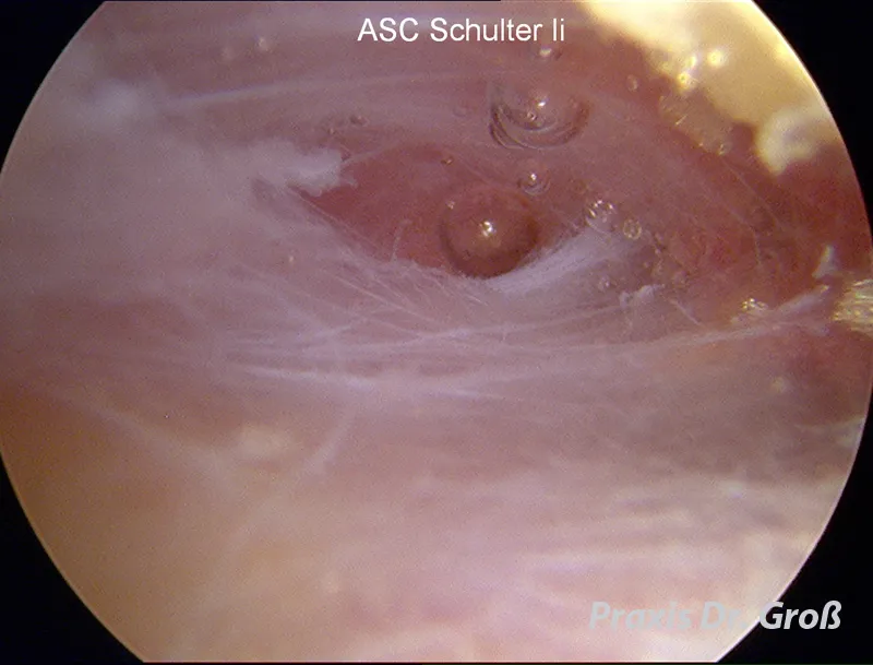
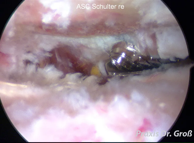
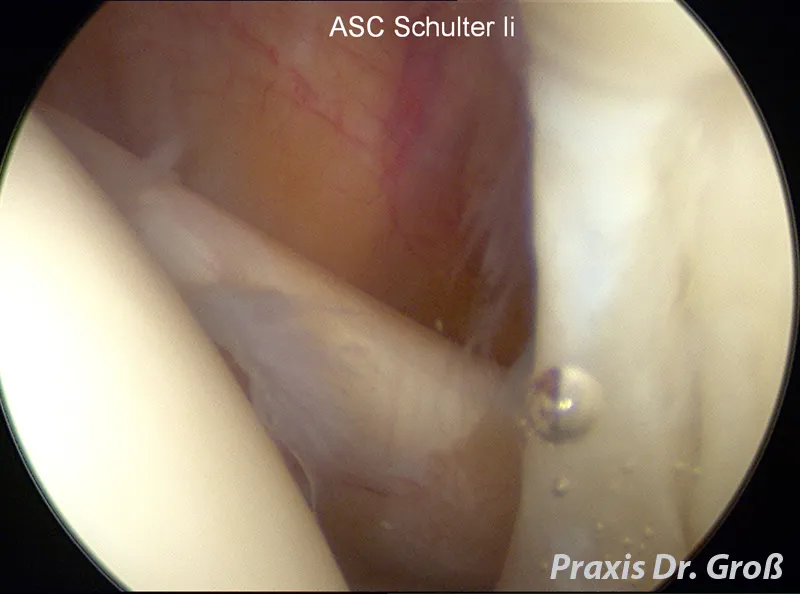
Images of a shoulder arthroscopy
Here you can see various images of a shoulder joint. The images were taken during a shoulder arthroscopy.
View from behind with the camera into a shoulder joint.
On the left side of the image, the humeral head is sectioned. The intact, smooth cartilage surface is clearly visible. Corresponding to the humeral head, on the right side of the image, is the acetabulum. The labrum, which consists of a strong connective tissue, is visible at the front edge of the bony acetabulum.
Also clearly visible in this image is a tendon ligament running from the right to the upper left. This tendon belongs to the subscapularis muscle, the anterior muscle of the rotator cuff.
A posterior view of the shoulder, between the rotator cuff tendon plate and the acromion. The camera is located in the subacromial bursa, which is slightly inflated by the irrigating fluid. The fine fibers of the bursa are clearly visible.
Posterior view of the shoulder between the rotator cuff tendon plate and the acromion. The chronically inflamed bursa was removed in this patient. The cause of the inflammation was an impingement syndrome with a large bony spur on the anterior edge of the acromion. This was a so-called type III acromion according to Bigliani.
To help the patient, this bony spur was milled from below using a motorized bone milling machine (acromionizer). The milling machine can be seen on the right side of the image. The processed bone surface of the acromion can be seen at the top of the image. The spur has already been completely removed.
This procedure required only two incisions, each approximately 0.5 cm long. This allowed healthy tissue structures to be preserved, resulting in less postoperative pain and faster wound healing. The operation was performed on an outpatient basis.