")
")
")
Shoulder Center Saar
The calcified shoulder
Cause and pain of calcific tendonitis, also called tendinosis calcarea.
Shoulder pain is common. In many cases, calcifications on or in the shoulder tendons are the cause of this shoulder pain. Patients diagnosed with calcific tendonitis, also known in the literature as tendinosis or calcific tendinitis, usually have symptoms for a long time before the diagnosis of calcific tendonitis is made.
You must have the Adobe Flash Player installed to view this player.
Zu Beginn der Erkrankung sind die Beschwerden meist noch gering und treten vor allem bei Überkopfarbeiten und Überkopf-Sportarten auf. Später treten die Beschwerden auch im Alltag auf und rauben Betroffenen nicht selten die Nachtruhe. Das Schlafen auf der Schulter wird in aller Regel zu Qual.
A new therapy, ultrasound-assisted microinvasive calcification removal, also known as needle lavage, promises fast and gentle relief. Numerous recent studies have already proven the effectiveness of this treatment with virtually no risks, leading shoulder specialists worldwide to recommend it. Unfortunately, it is still relatively unknown in Germany.
The exact cause of calcific tendonitis (calcific tendinosis) is not yet fully understood. Even the most recent scientific studies have not yet been able to definitively identify the triggers and processes within the tendon. However, there are many clues pointing to its cause. The most likely cause of calcification is reduced blood flow, resulting in a lack of oxygen (reduced partial pressure of oxygen in the tissue) in the tendon cells.
The tendons in the shoulder area naturally already have a poor blood supply. In some people, the blood vessels appear to be even less developed, which could then trigger calcification. Increased pressure on the tendon can also reduce blood flow to the tendon.
What increases the pressure on the shoulder tendons?
The most common causes of increased pressure on a shoulder tendon are muscular imbalance or shortening of the joint capsule. Muscles that work together unevenly lead to a minimal displacement of the humeral head. This phenomenon is not uncommon. A shortening of the joint capsule, usually in the posterior shoulder region, can also lead to this displacement of the humeral head. Due to our everyday lives, but also due to sports such as tennis, most people have significantly better developed chest muscles than their upper back and neck muscles. This alone results in muscular imbalance. Postural consistency in office work or monotonous activities, for example, does the rest.
The displacement of the humeral head narrows the space between the humeral head and the acromion (shoulder roof) or coracoid (a bony projection on the shoulder blade). This narrowing increases the pressure on the tendon and the associated blood vessels. They become compressed, thereby reducing blood flow. The result is the circulatory disorder already mentioned.
Recent studies show that extracellular matrix vesicles are responsible for calcification, which are stimulated to form calcium by the two factors mentioned above (pressure disturbance and lack of blood flow). However, calcification in the shoulder has nothing to do with calcium deposits in the blood vessels or "calcification" in the head.
In the literature, the findings regarding gender and age distribution vary considerably. However, many of these studies only survey a small patient population. Case numbers of 50 to 200 per study are not uncommon.
Due to the large number of patients in our practice over the last few years, we are able to publish our own scientific studies and evaluations on the subject of calcific tendonitis.
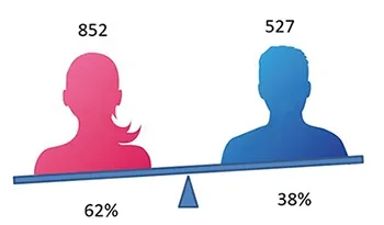
Our most recent analysis of more than 1,300 patients, conducted as part of my lectures, yielded the following results regarding gender and age distribution: Women are more frequently affected by calcific tendonitis than men.
Our study shows that women suffer from calcific shoulder significantly more often (approximately 60%) than men (approximately 40%).
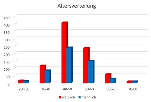
The age distribution of our study largely corresponds to the results of other scientific literature. Calcific tendonitis is predominantly a disease of middle age.
The condition is rare under the age of 30, and the incidence decreases with age. This also suggests that this condition is not a degenerative process. Otherwise, calcifications would become more common with age.
Typically, the disease calcific shoulder progresses in phases and describes a cycle
The four phases of calcific tendon disease - the starting and end point of the cycle is the healthy tendon
Precalcification
This phase is initiated by a lack of oxygen supply. It can be triggered by impaired blood flow or increased pressure in the tendon tissue with vascular compression.
In the affected area, tendon cells (tenocytes) transform into cartilage cells. This process is also known as chondroid metaplasia.
The cartilage cells now produce increased amounts of proteoglycans, a major component of cartilage. This leads to the formation of cartilage nests consisting of fibrocartilage within the tendon.
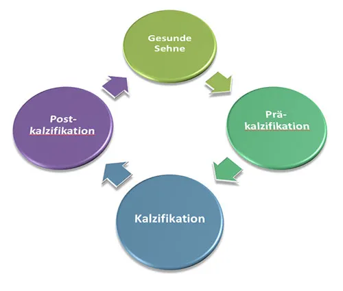
Calcification
During the calcification phase, calcium is deposited in the tendon. The cartilage cells formed in the previous phase deposit carbonate apatite, a calcium salt, around themselves. The remaining fibers in this area increasingly fuse until a calcification with a chalky and crumbly consistency forms.
This is followed by a resting phase during which the disease initially does not progress further. The calcification is enclosed by a firm fibrous capsule. Depending on the size of the calcification, patients describe restricted movement in the shoulder joint and occasional pain during this phase.
This resting phase is followed by resorption. For reasons not yet discovered, blood vessels and nerve fibers suddenly grow into the calcification. Scavenger cells (macrophages and rhinoserum cells) migrate into the calcification via the blood vessels and liquefy the calcification. However, this liquefaction causes the volume of the calcification to increase. Pressure within the calcification increases. This leads to irritation of pain fibers and the notorious pain of the disease.
A sudden increase in pressure can cause the calcium deposit to burst, and the now liquid calcium can pour into the overlying bursa. This leads to massive inflammation and often severe pain.
Postcalcification
This phase could also be called the healing phase. Fibroblasts (connective tissue cells) migrate into the defect area via the blood vessels. These form collagenous connective tissue to close the tendon defect.
In a so-called remodeling process, this collagen is gradually remodeled until intact tendon tissue is formed again
Healthy tendon
The last phase is also the starting point of the disease.
After the remodeling process, the tendon is healed again and we can speak of a healthy tendon.
The phases mentioned above demonstrate the natural progression of a calcified shoulder without any external influence. This means that even without the use of therapy, the natural progression usually leads to the calcium dissolving.
How can calcific shoulder be diagnosed?
The fastest, safest, and most gentle method is certainly ultrasound. Because this examination allows all tendon segments to be assessed by rotating the arm, calcifications can be detected very quickly, and their size and location can also be determined very accurately.
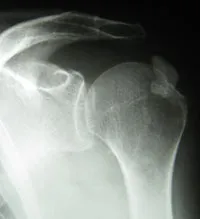
X-ray examinations can also clearly visualize calcifications. However, images from different angles are usually necessary to reliably visualize calcifications that are, for example, overlaid by the humeral head.
Detecting calcifications can be difficult with magnetic resonance imaging, so radiologists often recommend additional X-ray or ultrasound examinations.
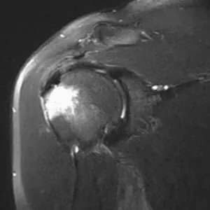
Above is an MRI image with a very nice representation of a calcification.
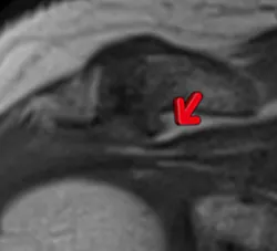
The magnified MRI image clearly shows the distinctive features of a calcified shoulder. The calcification is marked with the red arrow.
It's clearly visible that the calcification, shown in almost black, is located right in the middle of the tendon. Above and below the calcification, you can see the course of the tendon in gray.
What is typical for calcific tendonitis is that the calcification is not located on or under the tendon but in the middle of a tendon.
So why medical treatment?
The second phase of the disease, in particular, can last for a very long time, sometimes several years. For many patients, this situation leads to phases of recurring, severe pain with restricted movement, followed by phases of less severe discomfort. However, many patients complain of never being truly pain-free. For this reason, waiting for the calcification to resolve on its own is very unsatisfying for most patients.
If the calcium deposits dissolve spontaneously, acute pain often arises out of nowhere (this is also the typical symptom of acute calcium deposits in calcific tendinitis). This pain ranges from moderate shoulder pain to sometimes unbearable pain. Active movement of the shoulder is then no longer possible, and even light touch causes severe pain. In my experience, the faster a calcium deposit dissolves, the more severe the resulting inflammation and the more severe the pain. During this extremely painful phase, medical treatment is, of course, important.
When speaking generally about the treatment of calcific tendonitis, or tendinosis calcarea, it's important to understand that a doctor can't predict when or if the calcification will actually resolve completely. In the case of a painful accumulation of calcium in the shoulder tendon, it's important to determine which stage of the calcific tendon disease progression the patient is in. This is important for administering treatment appropriately.
The therapy options
In addition to open surgery, in which the calcification is removed under general anesthesia, the arthroscopic surgery, a keyhole procedure, has been established as another surgical procedure. Non-surgical treatment options include injections, painkillers, physiotherapy and Shock wave therapy A special diet or various nutritional supplements have not yet been shown to be effective in treating calcific tendonitis.
You must have the Adobe Flash Player installed to view this player.
Als neue sehr vielversprechende Therapie hat sich die microinvasive ultraschallgestützte Kalkentfernung (Nadel-Lavage) in lokaler Betäubung bereits bewährt. Dieses Verfahren wird bei entsprechender Indikation in unserer Praxis mit großem Erfolg durchgeführt. In den letzten 9 Jahren haben wir mehr als 4000 Behandlungen durchgeführt (Stand: 2.7.2017) und verfügen damit sicher über die meiste Erfahrung mit diesem Therapieverfahren Deutschland.
Neben der microinvasiven Therapie, der wir mittlerweile den Vorzug geben, sind wir auch in der Lage Kalkherde oder andere Erkrankungen der Schulter arthroskopisch zu beheben. Mit mehr als 350 jährlich durchgeführten Arthroskopien an der Schulter verfügen wir über eine sehr große Erfahrung und Routine bei diesen Eingriffen.
Als Schulterzentrum sind wir in der Lage Ihnen jedes der vorgenannten Verfahren zur Therapie der Kalkschulter anbieten zu können und sind nicht auf ein einziges Verfahren beschränkt.
Das nachfolgende Video zeigt eine arthroskopische Entfernung eines Kalkherdes:
You must have the Adobe Flash Player installed to view this player.